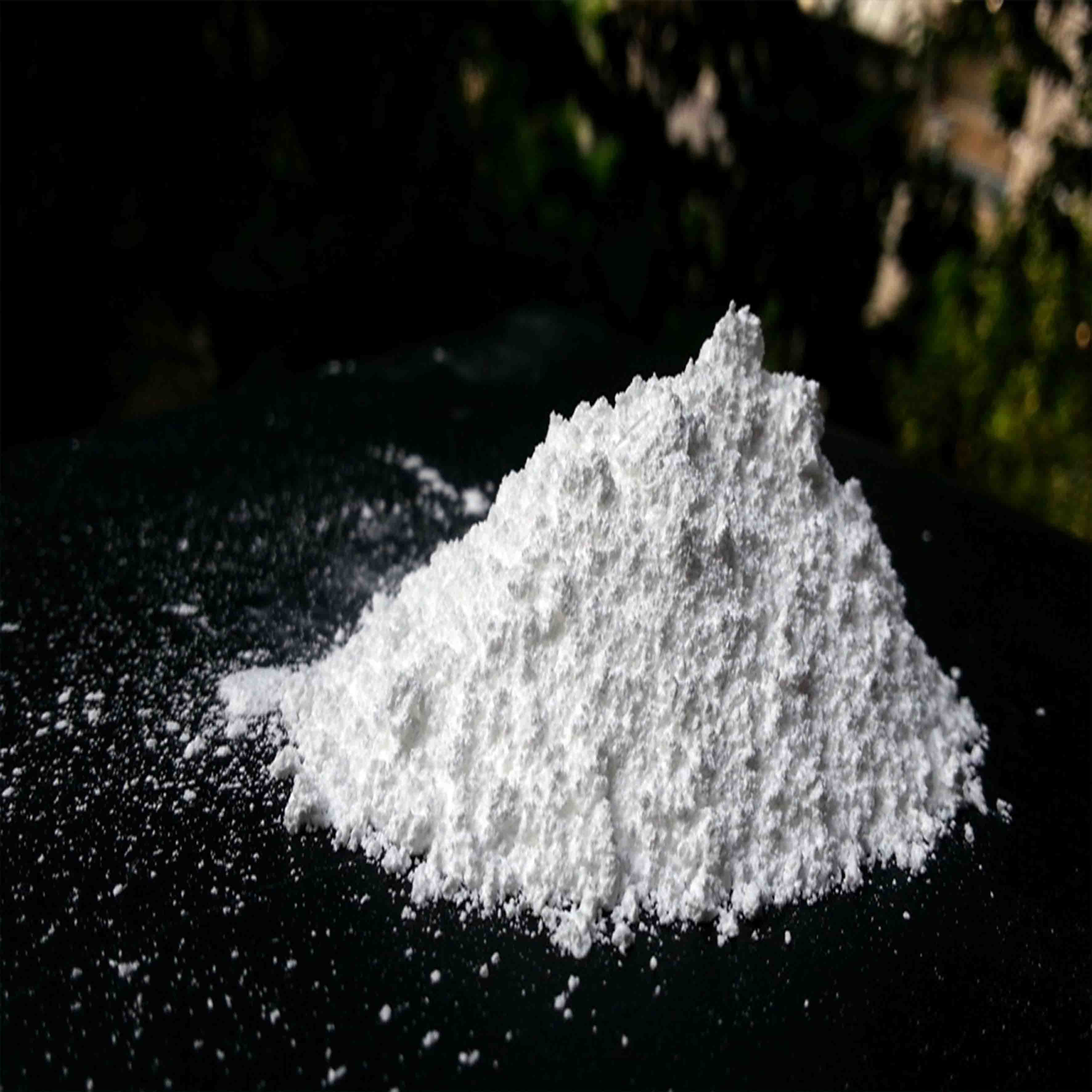
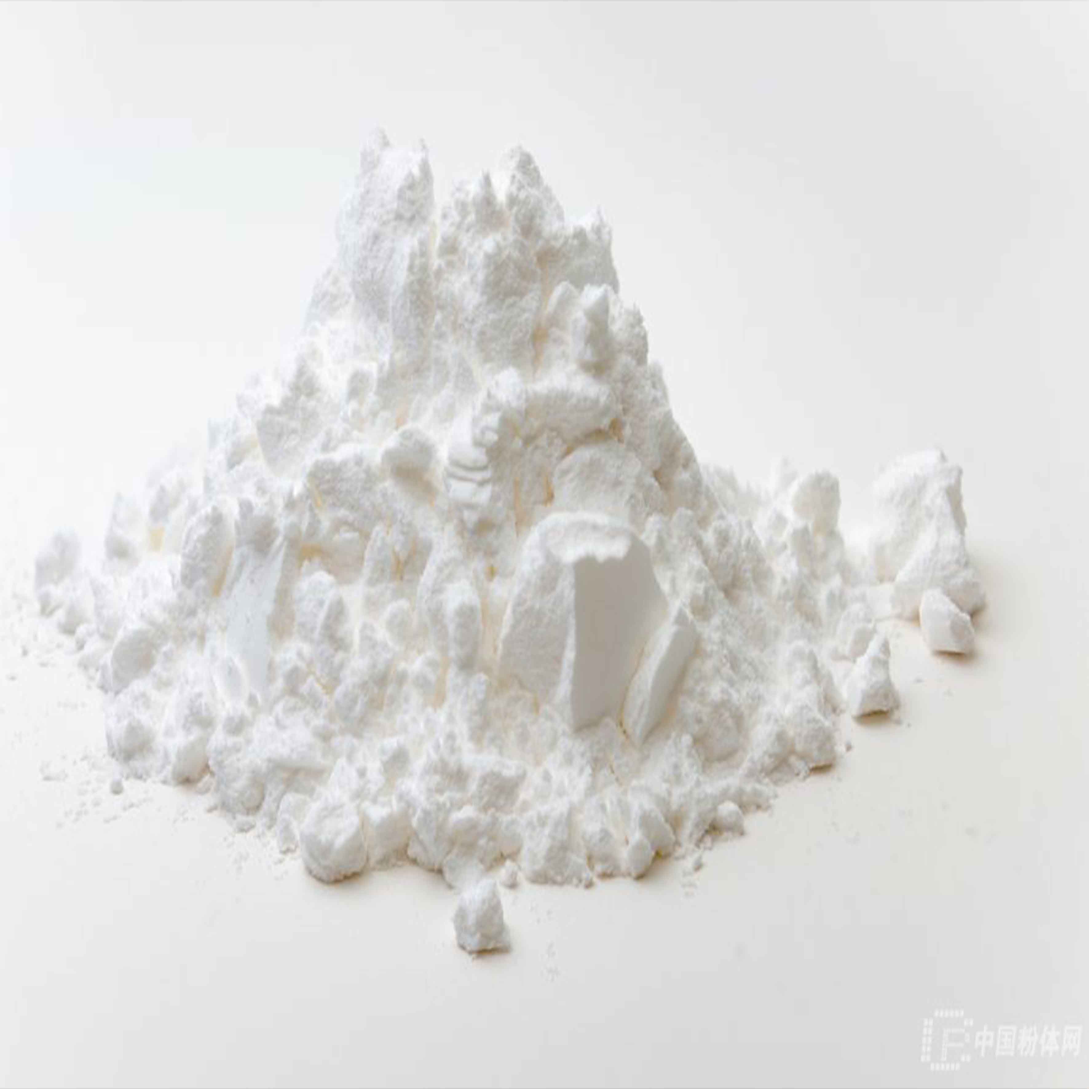
- The primary function of TiO2 in pigment production is its exceptional ability to provide brightness and opacity. When added to paints or coatings, it enhances their hiding power by reflecting light back to the observer's eye. This property not only improves the aesthetic appeal of the product but also reduces the amount of colorant needed, resulting in cost savings for manufacturers. Moreover, TiO2's high refractive index ensures that even small quantities can significantly impact the final appearance of the product.
It's sort of ironic, maybe ironic is the wrong word, that the ingredient in paint that makes your kitchen shiny also makes your Hostess cupcakes shiny, Environmental Working Group's senior vice president of government affairs Scott Faber added.
Now imagine the delicate skin on your face, on your children’s arms & legs. Each day un-knowingly, thinking we are doing the right thing, we slather them up with titanium dioxide in the form of sunscreen & send them out into the sun, all the while never knowing that once exposed to light titanium dioxide creates free radicals that are strong enough to damage steel roofing panels!!
Different dermal cell types have been reported to differ in their sensitivity to nano-sized TiO2 . Kiss et al. exposed human keratinocytes (HaCaT), human dermal fibroblast cells, sebaceous gland cells (SZ95) and primary human melanocytes to 9 nm-sized TiO2 particles at concentrations from 0.15 to 15 μg/cm2 for up to 4 days. The particles were detected in the cytoplasm and perinuclear region in fibroblasts and melanocytes, but not in kerati-nocytes or sebaceous cells. The uptake was associated with an increase in the intracellular Ca2+ concentration. A dose- and time-dependent decrease in cell proliferation was evident in all cell types, whereas in fibroblasts an increase in cell death via apoptosis has also been observed. Anatase TiO2 in 20–100 nm-sized form has been shown to be cytotoxic in mouse L929 fibroblasts. The decrease in cell viability was associated with an increase in the production of ROS and the depletion of glutathione. The particles were internalized and detected within lysosomes. In human keratinocytes exposed for 24 h to non-illuminated, 7 nm-sized anatase TiO2, a cluster analysis of the gene expression revealed that genes involved in the “inflammatory response” and “cell adhesion”, but not those involved in “oxidative stress” and “apoptosis”, were up-regulated. The results suggest that non-illuminated TiO2 particles have no significant impact on ROS-associated oxidative damage, but affect the cell-matrix adhesion in keratinocytes in extracellular matrix remodelling. In human keratinocytes, Kocbek et al. investigated the adverse effects of 25 nm-sized anatase TiO2 (5 and 10 μg/ml) after 3 months of exposure and found no changes in the cell growth and morphology, mitochondrial function and cell cycle distribution. The only change was a larger number of nanotubular intracellular connections in TiO2-exposed cells compared to non-exposed cells. Although the authors proposed that this change may indicate a cellular transformation, the significance of this finding is not clear. On the other hand, Dunford et al. studied the genotoxicity of UV-irradiated TiO2 extracted from sunscreen lotions, and reported severe damage to plasmid and nuclear DNA in human fibroblasts. Manitol (antioxidant) prevented DNA damage, implying that the genotoxicity was mediated by ROS.
 Anatase titanium dioxide is typically produced by the chloride process, which involves the chlorination of titanium ore to produce titanium tetrachloride Anatase titanium dioxide is typically produced by the chloride process, which involves the chlorination of titanium ore to produce titanium tetrachloride

 The device comes with a user-friendly interface that makes it simple to set up and manage The device comes with a user-friendly interface that makes it simple to set up and manage